Vitreoretinal surgery in Jalandhar, Phagwara, Punjab

Vitreoretinal surgery in Jalandhar, Phagwara, Punjab, Mitra Eye Hospital under the supervision of Dr. Harinder Mitra is fully equipped with state-of-the-art Vitreo Retinal Surgical Treatment. This surgical approach is performed under various issues linked to retinal problems. The 3D Genuinity visualization system helps the surgeon to perform the heads-up vitreoretinal surgery. Vitreoretinal surgery Cost in Punjab starts from Rs 20,000 and it can range high depending on your condition.
Expert Eye Care, Latest Eye Treatment
What is Vitreoretinal Surgery?
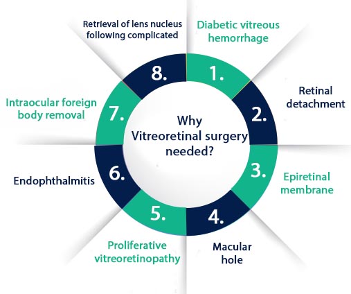
Vitreoretinal Eye Surgery is performed under the group of latest procedures to treat the inside of the eyes with conventional or laser surgical instruments. Under the supervision of Dr. Harinder Mitra, Dr. Akshay Mitra and his expert team will perform this delicate surgery on the part where there is gel-like vitreous and retina (light-sensitive membrane) are found. This approach is beneficial for different eye conditions which are linked to age-related macular degeneration, macular hole, detached retina, CMV retinitis, epiretinal membrane, and diabetic vitreous hemorrhage.
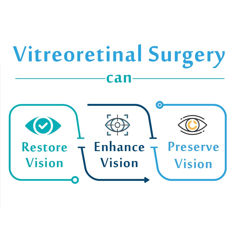
| Do you know what a vitreoretinal surgical procedure can do? | ||
| Restore Vision | Preserve Vision | Enhance Vision |

What are the most common reasons to perform vitreoretinal eye surgery?

- Diabetic retinopathy: Diabetic patients will have complications like blood vessels getting damaged in the retina.
- Floaters and flashes: Flashes occur when vitreous moves in the eyes and then pulls the retina which creates a light flash. Floaters are seen when substances are formed in the retinal tear, vitreous, or hemorrhage.
- Macular holes: It triggers due to age in which the vitreous shrink and retina pulls & tears a hole in the section which is known as the macula. This can affect the vision.
- Retinal detachment or tears: Retina separation or tears occur in the back part of the eye. Patients will experience a sensation that closes on the peripheral vision.
- Macular pucker: The retina will have a small area as wrinkled which affects the focus and leads to distorted vision.
- Retinitis pigmentosa: A rare genetic problem that affects the retina cells and might lead to vision loss (extreme). It will have symptoms like peripheral vision loss, central vision loss, and night vision loss.
- Retinopathy of prematurity: Its occurrence is mostly in premature babies as the retina is not developed completely and it leads to abnormal blood vessels which lead to retina detachment & distortion.
- Retinoblastoma: One of the types of eye cancer occurs in infants or early childhood.
Take Utmost Care and Prevent the chances of Permanent Vision Loss
How does the Vitrectomy procedure work?
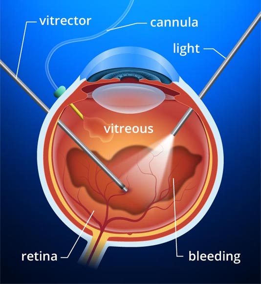
During the surgery, the gel-like substance or vitreous humor is removed. Undergoing this approach will solve the vision issues when the foreign matter affects the pristine area which is the inner part of the eyes. One of the foreign substances in the blood that can occur due to diabetic vitreous hemorrhage.
Dr. Harinder Mitra, “With vitrectomy the patient’s vision is restored who have diabetic retinopathy. We take out the natural vitreous which has become clouded due to blood vessel leakage and instead clear fluid is added.”
Once the vitreous humor is taken away and the entire area is cleaned. After that, the saline liquid is injected to replace it with the vitreous humor
Once the surgeon removes the vitreous humor and clears the area, he or she usually injects a saline liquid to replace the vitreous humor that ordinarily fills up the inner chambers of the eye which fills in the inner part of the eyes.
Is the vitrectomy procedure done under anesthesia?
In most cases, the patient needs general anesthesia and sometimes local anesthesia is used. The surgeon will create tiny incisions (around 3) to create the eye-opening and through the opening instruments are added. Inside the incisions, the doctor will place the instruments which include:
- Light pipe: It serves as a microscope that has a high-intensity flashlight to use within the eyes.
- Infusion port: Helps to replace the fluid present in the eye and put saline solution. Through this, eye pressure will be proper.
- Vitrector, or cutting device: Through this, the eye’s vitreous gel is removed and everything is controlled properly. This way, the vitreous humor is removed.
What to expect after a Vitrectomy?
- There are different things involved with the surgery and only the surgeon who understands your condition will give you the best results.
- Following the procedure, you will use antibiotic eye drops for the initial week. The doctor will prescribe you eye drops that need to be used for several weeks.
- Make sure to follow the surgeon’s advice carefully. You will be able to see the final results after a few weeks. All in all, you need to follow all the instructions given by your ophthalmologist.
Epiretinal Membrane Peeling (Membranectomy)
Epiretinal membrane (ERM) is also referred to as macular pucker and cellophane retinopathy includes membrane growth which is the same as that of scar tissue around the macula. Many times it happens that the macula, the sensitive part of the retina responsible for the visual activity and color vision, encounters growth of a scar across the retina, causing central vision distortion. The solution to the problem is Membranectomy, also known as macular pucker or cellophane retinopathy. After completing the process of vitrectomy, the surgeon making use of precise, sharp forceps will peel away the membrane from the retina. Tiny sutures seal the incisions that do not require removal. The recovery rate from the membrane to my is high in many cases, yet exceptions prevail. It does not carry many risks with it. However, not following the precautionary measures can lead to infection and increase the time of recovery.
|
Reasons to get membranectomy |
|
| Epiretinal Membrane is present | Problem of Vision Distortion or reduced vision due to ERM. |
How does the membrane peeling procedure work?
Initially, the procedure starts with a vitrectomy. The surgeon uses the fine forceps under high magnification to gasp and feel the membrane from the retina. The membrane is removed by the diamond-dusted instruments. Our surgeon performs the surgery with utmost precision to make sure eyes are not affected.
Surgery For Proliferative Vitreoretinopathy
Following retinal detachment, the patient will have the most common complication named Proliferative vitreoretinopathy (PVR). The doctor will diagnose it and then perform the surgery.
With time the retinal tissue becomes rigid, hard, and immobile increasing visionary and treatment problems. Ironically this problem has a reverse relation to the treatment. In other words, during the developmental stage, the retina is compliant, and PVR at this stage is not possible as the problem lacks clarity.
The retina is neither in its exact form nor completely deformed in shape, location. Over the period, the retina becomes detached, rigid taking the white, fibrotic appearance increasing the chances of removal. Vitrectomy follows the filling of gases and liquids into the eye to help keep the retina reattached to the outer walls of the eye.
The process many times also calls in need for sclera. Artificial material is inserted into the eye to maintain the pressure that helps in keeping the retina attached to the wall. The results of the process enable Ambulatory vision, but the precise and sharp clarity in vision remains far from reality.
Recovery after surgery for PVR – Takes time
50% of the patient will have a vision but that is to see the objects within a close range. Although, the reading vision will be low.


